
ColonView™

AI-assisted CT analysis for colorectal imaging — bowel preparation assessment, polyp detection, segmentation & measurement.

Investigational software. Development and validation ongoing.
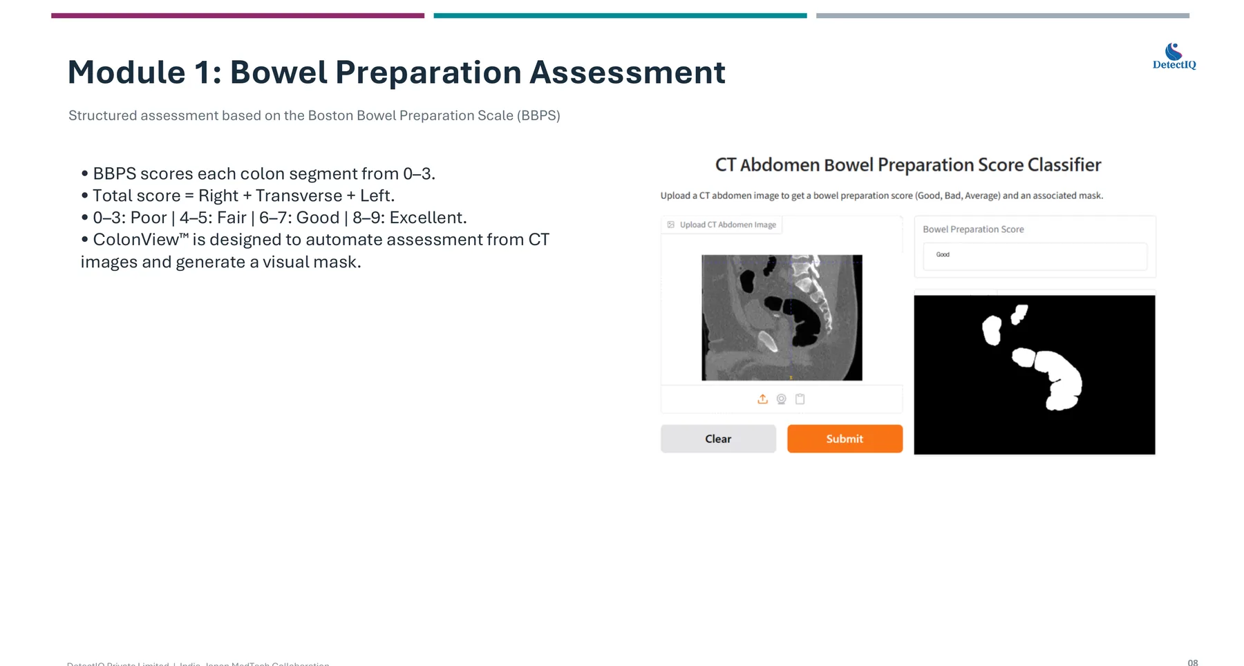
CT Abdomen Bowel Preparation Score Classifier interface with segmentation mask
Module 1 — CT Abdomen Bowel Preparation Score Classifier: CT input with colon segmentation mask
Three modules, one workflow

Module 1 — Bowel Preparation Assessment

Structured assessment based on the Boston Bowel Preparation Scale (BBPS). BBPS scores each colon segment 0–3; total score = Right + Transverse + Left.
  • 0–3 Poor · 4–5 Fair · 6–7 Good · 8–9 Excellent
  • ColonView™ automates this assessment from CT images
  • Generates a visual mask of the colon regions
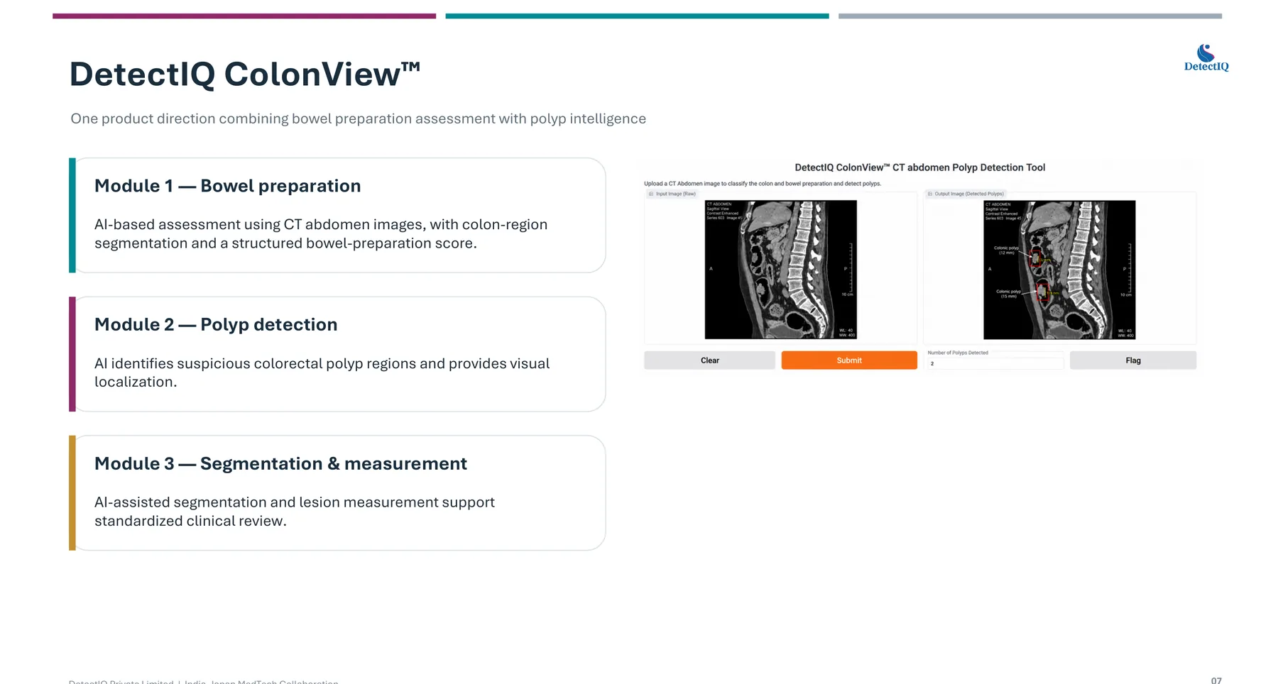
CT Polyp Detection Tool interface
Module 2 — CT Polyp Detection Tool: suspicious regions with visual localization

Module 2 — Polyp Detection

The AI identifies suspicious colorectal regions — polyps, masses, abnormal wall thickening and other suspicious findings — and provides visual localization with overlays for clinician review.
The AI does not need to “be the doctor.” Its job is to help the clinician notice, localize and assess findings more efficiently.
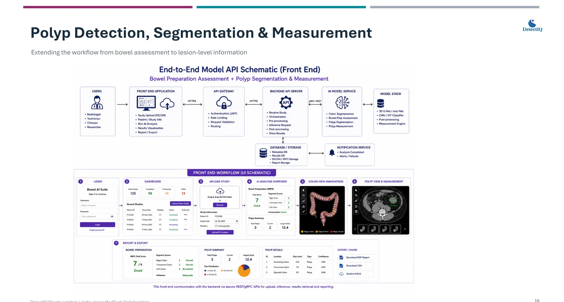
Clinical dashboard: colon navigation view and polyp measurement view
Module 3 — Clinical dashboard: colon navigation and lesion measurement views

Module 3 — Segmentation & Measurement

AI-assisted segmentation and lesion measurement support standardized clinical review:

  • Location of each finding
  • Size in mm and size distribution
  • Confidence per finding
  • Report-ready structured output
Reference

The scale we automate — BBPS

Each segment (right, transverse, left) is scored 0–3; the total is the sum of the three segments.

Total BBPSInterpretation
0–3Poor
4–5Fair
6–7Good
8–9Excellent
The problem

Why CT-based detection is hard

Small & flat lesions

Polyps below 5 mm are harder to detect; flat lesions can produce minimal protrusion from the bowel wall.

Inadequate bowel preparation

Residual stool and fluid can mimic polyps, hide actual polyps and reduce image quality.

Poor bowel distension

Folded or collapsed bowel segments can obscure lesions and make small abnormalities difficult to evaluate.

Partial-volume effects

Small lesions occupying only part of a CT slice may blend into surrounding tissue or appear smaller.

Integration

Designed to complement existing imaging workflows

  • Input: routine abdominal CT in DICOM format
  • Integration: PACS/DICOM-compatible, with the option of a standalone clinical tool
  • Output: visual review overlays, structured findings and report-ready measurements

Fits within the radiologist's existing workflow — no additional imaging procedure required.
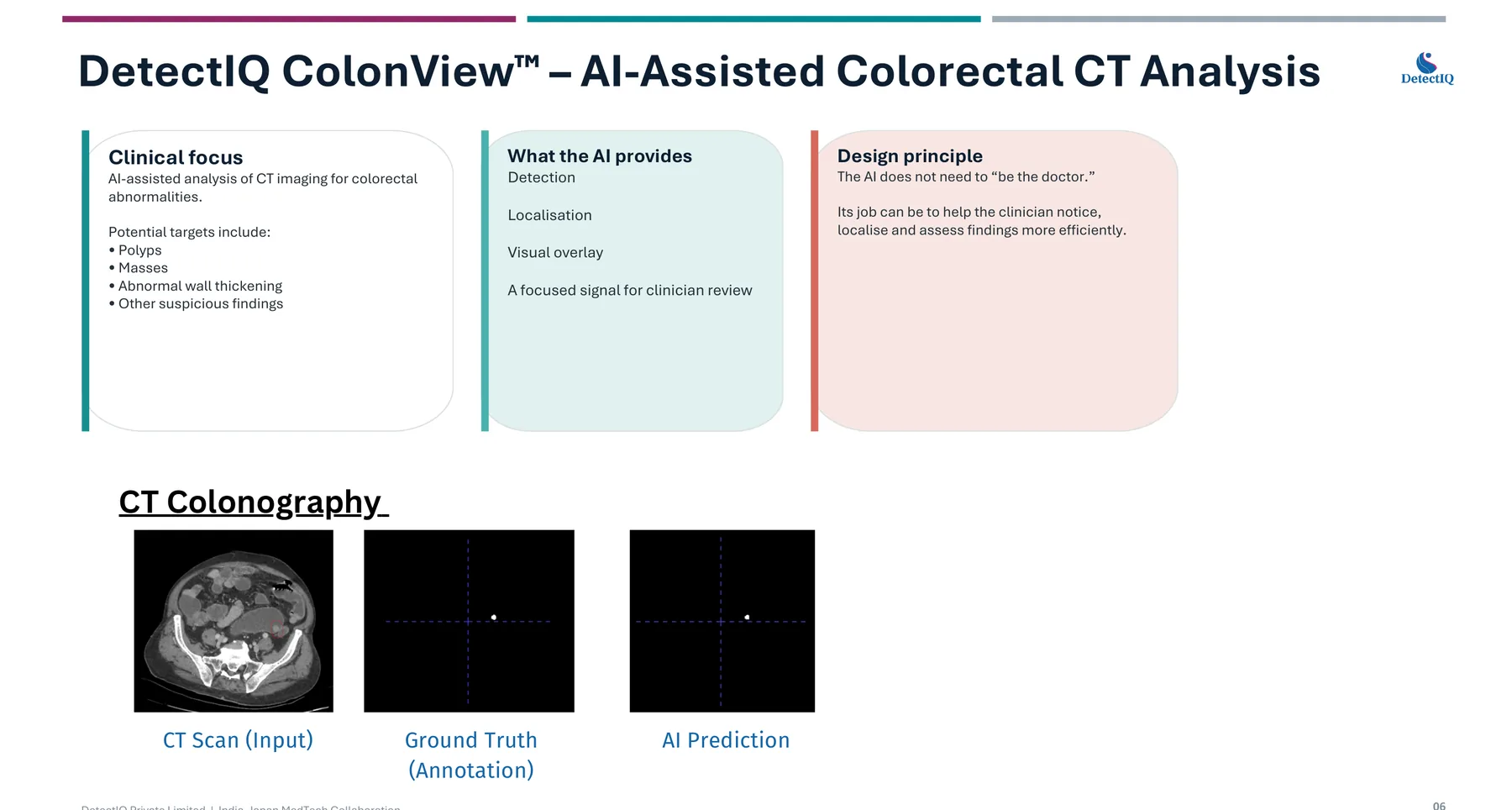
Ground truth annotation versus AI prediction comparison
Ground truth (annotation) vs. AI prediction — visual comparison from development
Stage

Where we are today

Research ✓
Prototype ✓
Validation (current)
Pilot
Commercial

Current focus: clinical evaluation and product refinement. Next step: evaluation on external (e.g. Japanese) datasets and workflows. Longer term: integration and commercialization.

A live demonstration of the tool interface is available on request; validation datasets and detailed performance results are shared under NDA.

ColonView™ is investigational software under development for research and clinical-validation purposes. It is not a medical device, has not been approved or cleared by CDSCO (India), PMDA (Japan) or any other regulatory authority, and must not be used as the basis for diagnostic or treatment decisions.